What is the Function of the Nervous System?

Hello, and welcome to this Mometrix video covering the nervous system!
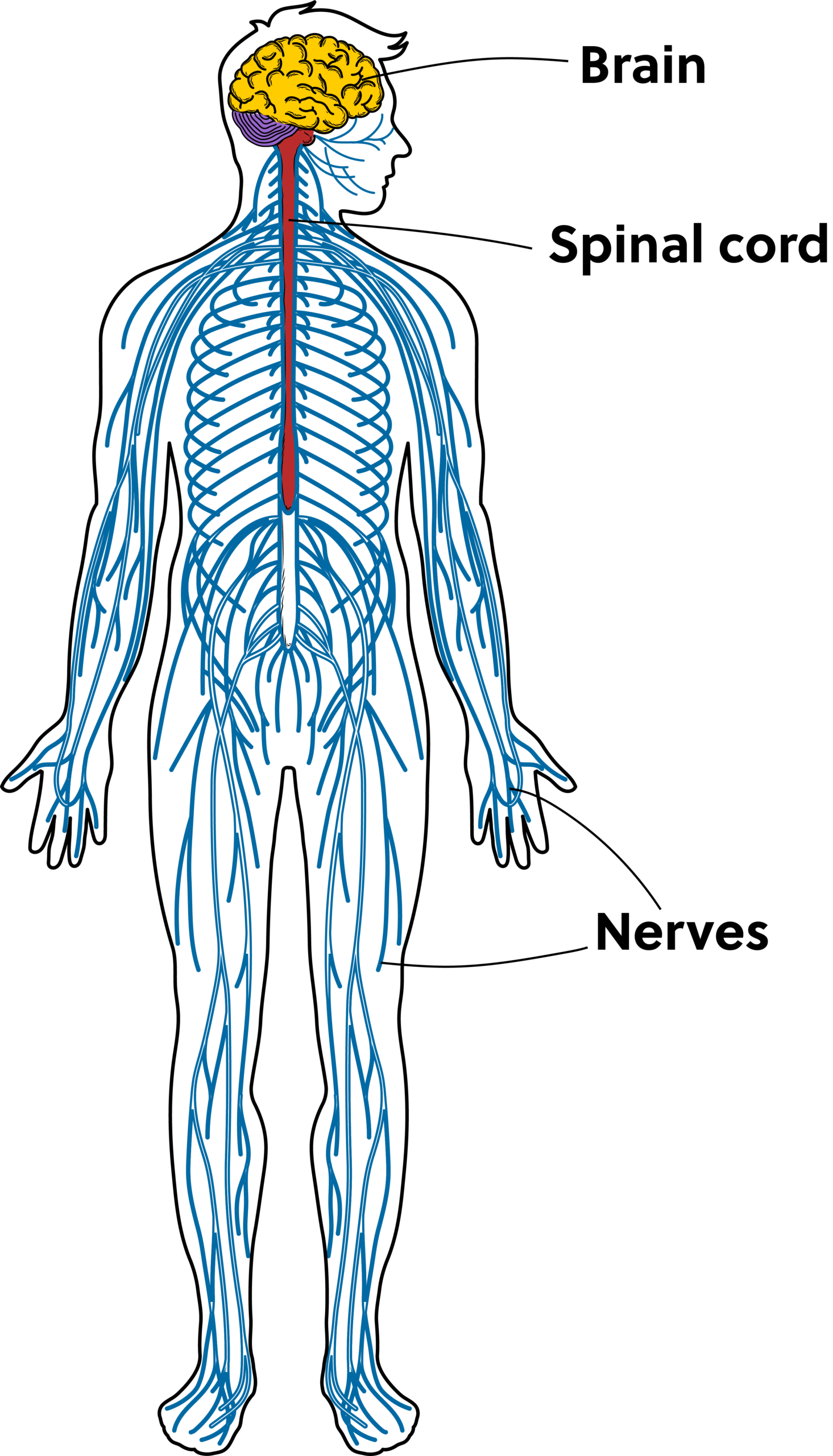
All systems of the body are important, but the nervous system is the system that has an impact on EVERYTHING the body does. It’s responsible for getting us to react to changes both inside and outside of our bodies, and it’s the reason different parts of our body can communicate with each other. It includes our brain and spine as well as nerves throughout the body.
The nervous system has three main tasks.
- First, it collects and receives sensory information from both inside and outside the body.
- Second, it processes the sensory data.
- Third, it then produces a response in the relevant part of the body.
Today, we are going to take a closer look at how various aspects of the nervous system help us accomplish these tasks.
The nervous system is separated into two main divisions: the central nervous system, which includes the brain and the spinal cord, and the peripheral nervous system, which includes the nerves that stem from the central nervous system and spread throughout the body.
First, let’s take a look at the central nervous system.
Central Nervous System (CNS)
The main function of the central nervous system is to receive and integrate sensory data and then stimulate a response within the body. Like we mentioned earlier, it is composed of the brain and the spinal cord.
The brain is composed of four main regions: the cerebrum, cerebellum, brainstem, and diencephalon.
Cerebrum
The cerebrum is the largest part and sits along the top and side portions of the brain. It is separated into two distinct halves: the left and right hemispheres. Interestingly enough, the left hemisphere controls the right side of the body, and the right hemisphere controls the left side of the body. This is referred to as contralateral control.
The cerebrum is also made up of four different lobes: the frontal lobe, the temporal lobe, the parietal lobe, and the occipital lobe.

The frontal lobe is (surprise surprise) at the front of the brain. It is responsible for consciousness and other higher-level cognitive functions, including problem-solving, planning, memory, and communication. It also contains the primary motor cortex, which initiates body movement.
The brain has two temporal lobes, one in each hemisphere located near your ears. Their primary functions are to process auditory stimuli, store memories, regulate emotions, and help us to communicate language.
The parietal lobe is located at the top of the head behind the frontal lobe. This lobe processes sensory input related to touch, temperature, and pain.
The occipital lobe sits in the back of the cerebral cortex, and its primary role is to process visual information.
Cerebellum
The next region of the brain, the cerebellum, is located at the back of the brain. It helps coordinate our movements and is responsible for balance, posture, and muscle memory.
Brainstem
The third region of the brain, the brainstem, is located at the base of the brain and is the connection between the brain and the spinal cord. It is responsible for many of the basic vital functions required for life.
The brainstem has three primary parts: the midbrain, the pons, and the medulla.
The midbrain is the top section of the brainstem. It plays an important role in the regulation of our eye movements.
The pons is the second section of the brainstem, and it is the connection point between the brain and the spinal cord. The pons plays an important role in the regulation of our sleep-wake cycles, our ability to perceive pain, and our ability to move our face.
The medulla sits at the bottom of the brainstem. It is responsible for crucial life-sustaining tasks such as regulating respirations, heart rate, and blood pressure.
Diencephalon
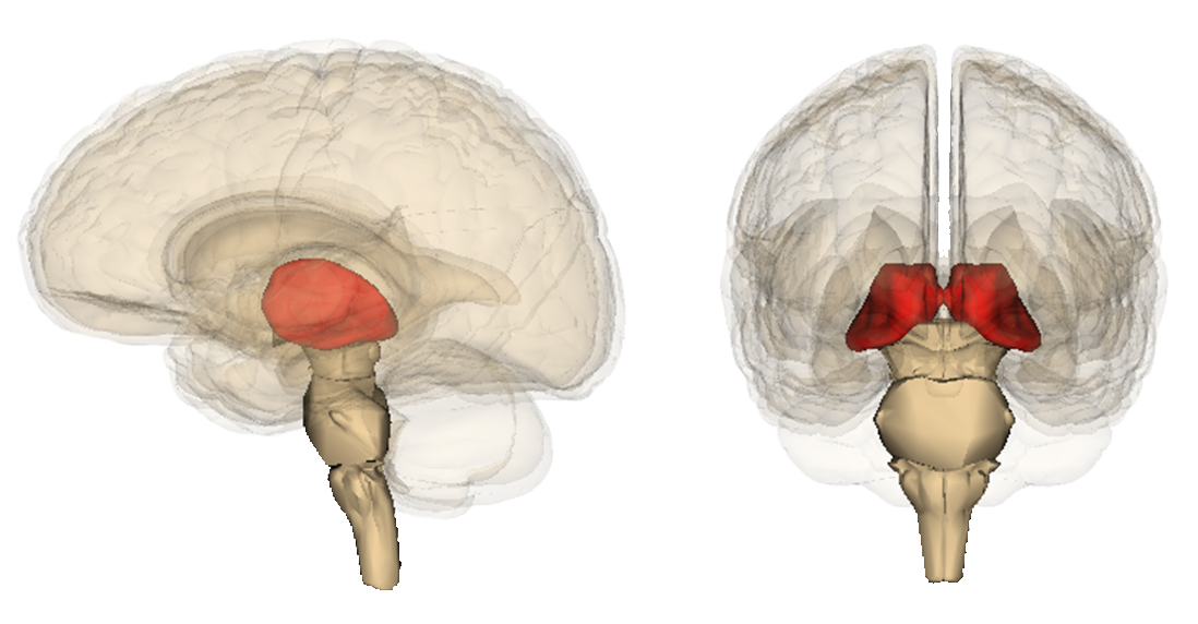
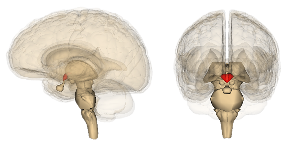
The fourth region of the brain, the diencephalon, is located deep within the brain above the brainstem and beneath the cerebral cortex. It is sometimes referred to as the interbrain, and it contains several important features, including the thalamus and hypothalamus.
The thalamus is responsible for initially processing nearly every sense experienced by the body. It then relays the sensory data to the appropriate part of the brain for further processing.
The hypothalamus is a key link between the nervous system and endocrine system, and it helps our body maintain homeostasis.
Spinal Cord
The second primary component of the central nervous system is the spinal cord. The spinal cord runs within the length of the spinal column, which consists of 33 vertebral bones meant to protect the spinal cord.
The spinal cord functions as a two-way channel between the body and the brain. Motor data from the brain travels down the spinal cord and out to the body. In the reverse direction, sensory data from the body travels back up the spinal cord to the brain.
Peripheral Nervous System (PNS)
Now, let’s move onto the second division of the nervous system, the peripheral nervous system. The peripheral nervous system is responsible for communication between your central nervous system and the rest of your body. It carries sensory data from your body back to your brain, and it brings motor impulses from your brain to the target organ, muscle, or gland.
The peripheral nervous system is made up of nerves that stem from the brain and spine. This includes 12 cranial nerves and 31 pairs of spinal nerves.
The basic unit of the peripheral nervous system is a nerve cell, called a neuron, which has several features that allow it to relay messages rapidly throughout the body.
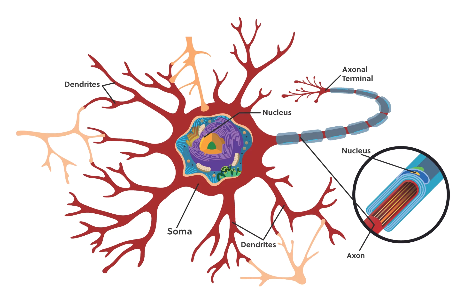
The main cell body is called the soma. Branching off the soma is a long nerve fiber called the axon, which carries nerve signals away from the cell body.
There are also many smaller fibers branching off the soma, called dendrites, and these receive signals from other neurons. “Ganglia” refers to clusters of neuron cell bodies, whereas a “nerve” refers to clusters of axon fibers.
Within the peripheral nervous system, there are two sub-systems: the somatic nervous system and the autonomic nervous system.
Somatic Nervous System (SNS)
The somatic nervous system involves aspects of the nervous system that we are consciously aware of and can control. It contains sensory neurons, which carry information from our senses—what we see, touch, and hear—to our brain. And, in the opposite direction, it contains motor neurons, which carry commands from the brain to our muscles to initiate voluntary bodily movements and involuntary reflexes.
Autonomic Nervous System (ANS)
The second sub-system of the peripheral nervous system is the autonomic nervous system. This system manages involuntary movements and functions, such as communication with our internal organs and digestion.
There are two divisions within the autonomic nervous system: the sympathetic nervous system and the parasympathetic nervous system.
The sympathetic nervous system is the system that responds to threats and activates our “fight or flight” response. This system increases our level of alertness, speeds up our heart rate, causes muscle contraction, and shuts off any function that is not essential for survival.
The second division within the autonomic nervous system, the parasympathetic nervous system, does the opposite. It activates our body’s “rest and digest” state and manages the body’s function while at rest. It relaxes our muscles, slows our heart rate, and restores everything to a state of relaxation and balance.
Review
We just covered a lot of information. Let’s summarize what we’ve reviewed with a very generalized diagram of the nervous system so we can see how it is all connected.
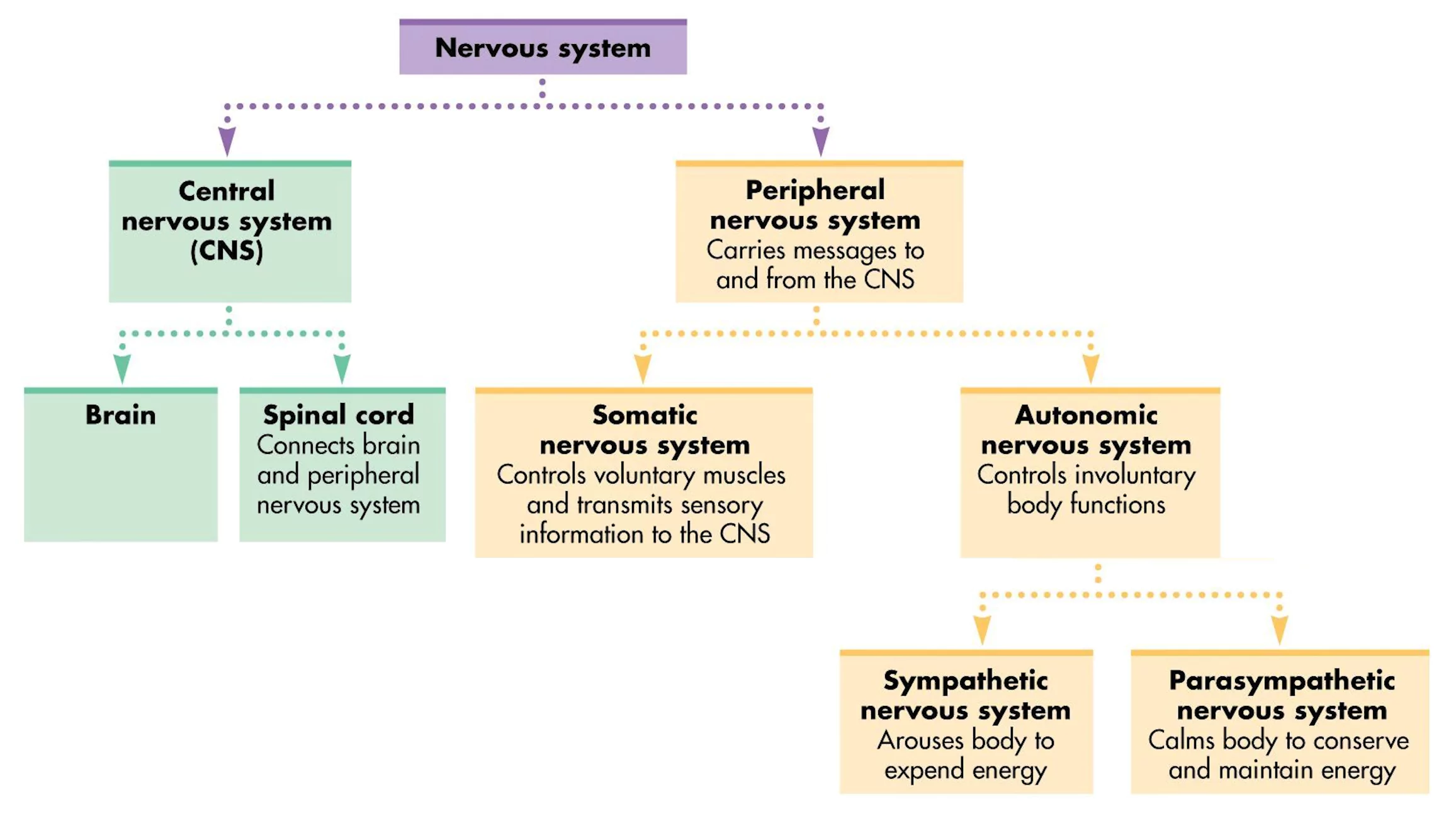
Within the nervous system, there are two main divisions: the central nervous system (which includes the brain and spinal cord that receive and process sensory data) and the peripheral nervous system.
The peripheral nervous system then has two subdivisions: the somatic nervous system (which controls voluntary movements and reflexes) and the autonomic nervous system (which controls involuntary movements).
Within the autonomic nervous system, there is the sympathetic nervous system (which controls our “fight or flight” response) and the parasympathetic nervous system (which controls our “rest and digest” response).
All of these systems work together to help you understand and react to the world around you.
I hope this overview of the nervous system has been helpful to you. Thanks for watching, and happy studying!
Frequently Asked Questions
Q
What is the function of the nervous system?
A
The main function of the nervous system is to coordinate the activities of the body by sensing, interpreting, and issuing commands as a response to conditions in the body’s environment.
Q
How does the nervous system work?
A
Messages are sent across the plasma membrane of neurons through a process called action potential. These messages occur when a neuron is stimulated past a necessary threshold. These stimulations occur in a sequence from the stimulation point of one neuron to its contact with another neuron. At the point of contact, chemical synapse, a substance is released that stimulates or inhibits the action of the adjoining cell. A network of nerves composed of neurons fans out across the body and forms the framework for the nervous system. The direction the information flows depends on the specific organizations of nerve circuits and pathways.
Q
What organs are in the nervous system?
A
The brain and spinal cord make up your central nervous system.
Q
How does the nervous system maintain homeostasis?
A
Homeostasis is the regulation of internal chemistry to maintain a constant internal environment. This state is controlled through various feedback mechanisms that consist of receptors, an integrator, and effectors. Receptors such as mechanoreceptors or thermoreceptors in the skin detect the stimuli. The integrator such as the brain or spinal cord receives the information concerning the stimuli and sends out signals to other parts of the body. The effectors such as muscles or glands respond to the stimulus. Basically, the receptors receive the stimuli and notify the integrator, which signals the effectors to respond.
Q
What effect does exercise have on the nervous system?
A
Moderate exercise can improve mood and help cognitive function.
