
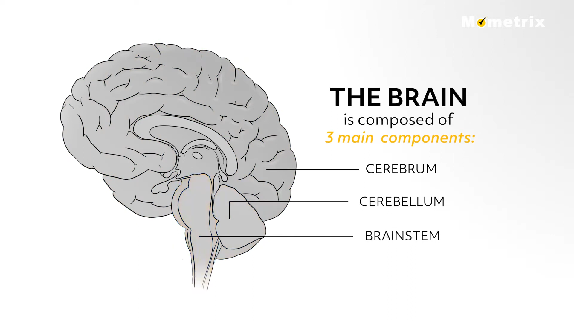
In today’s video we are going to talk about the anatomy of the brain and the corresponding functions. The brain is composed of three main components— the cerebrum, the cerebellum, and the brainstem. Each have their own distinct features and functions that we will be discussing in this video. Be sure to check out our other anatomy videos for quick overviews of the body systems and their actions on the body.
Cerebrum
The cerebrum is the largest part of the brain and is responsible for vision, hearing, interpreting speech, and dealing with emotions.
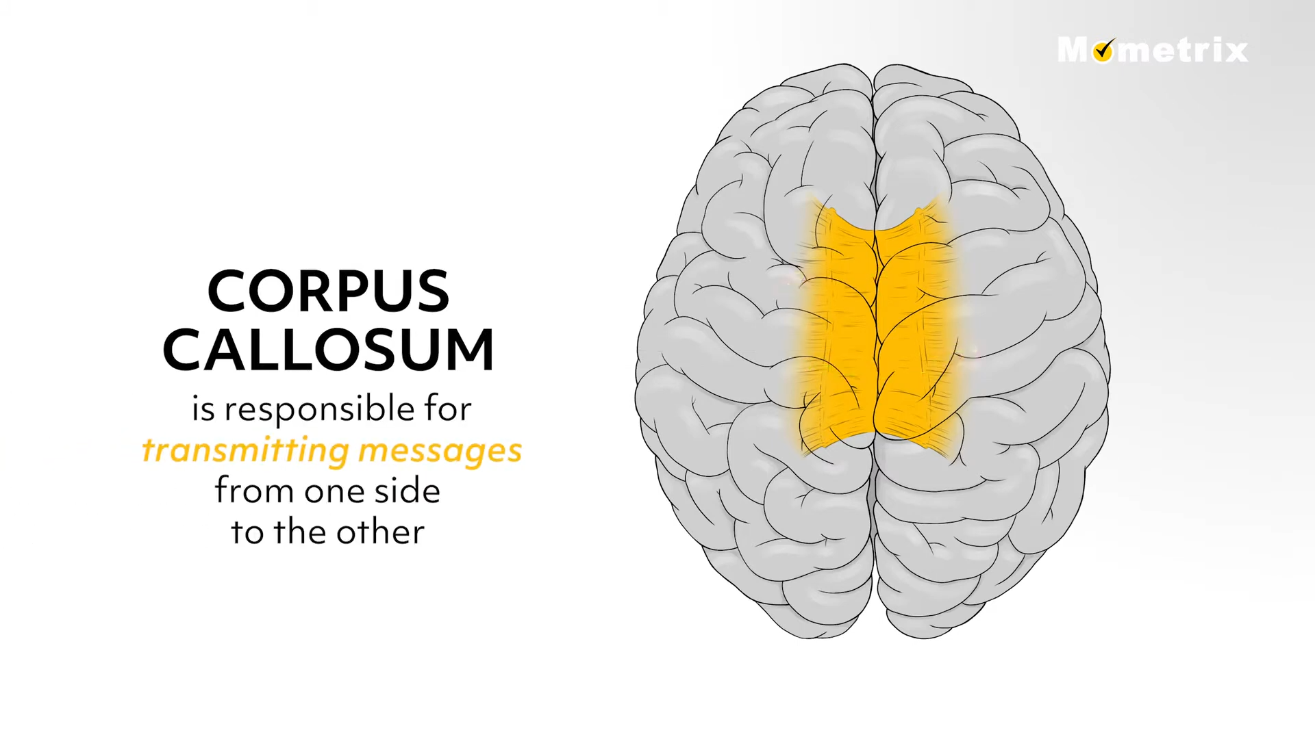
The cerebrum is divided into the right and left hemispheres and is joined in the middle by a bundle of fibers called the corpus callosum. The corpus callosum is responsible for transmitting messages from one side to the other.
Each hemisphere controls the opposite side of the body. For example, if a stroke-like event happened in the left side of your brain, the extremities on your right side would be affected. The right hemisphere is responsible for creativity, musical skills, and artistic abilities.
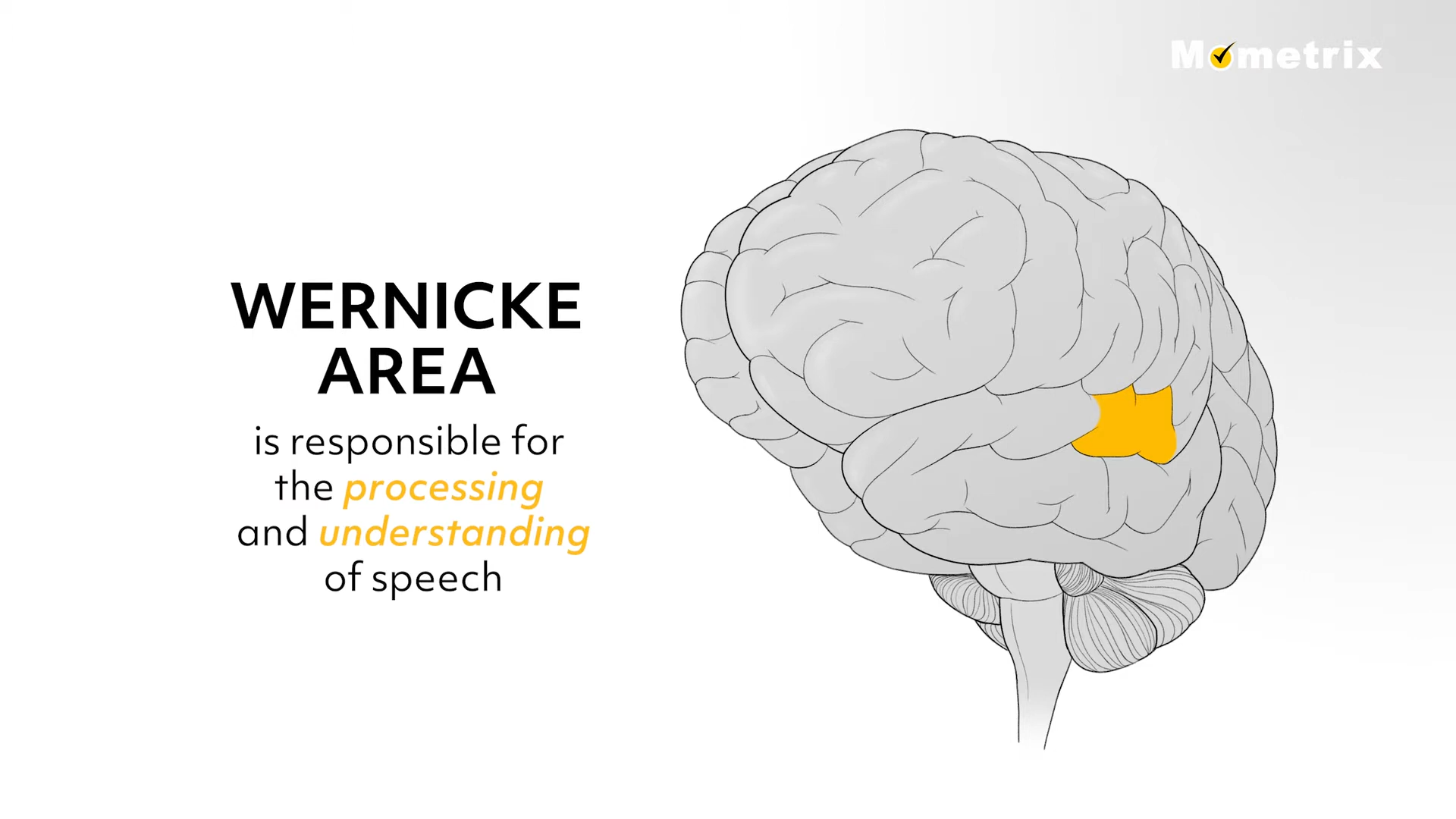
The left hemisphere controls comprehension, writing, speech and language and is usually referred to as the “dominant” hemisphere. There are two special areas in the left hemisphere that help the brain decipher and process speech. The Broca area of the brain is responsible for speaking and writing. Consequently, if this area is damaged, a person can still understand what is being spoken but will have difficulty in speaking or writing. The Wernicke area of the brain is responsible for the processing and understanding of speech, so if this area is damaged, one could speak or write but the words would not usually make sense or have any meaning.
The cerebrum is also divided into four lobes: the parietal lobe, the occipital lobe, the frontal lobe, and the temporal lobe. The frontal lobe is in the anterior portion of the cerebral cortex, generally in the “forehead” area. It is the largest brain lobe and is responsible for motor functioning, memory recall, impulse control, language and speech, reasoning, and judgement. Damage to this area of the brain can have a profound effect on speech and language, fine motor skills, personality changes, and thinking difficulties.
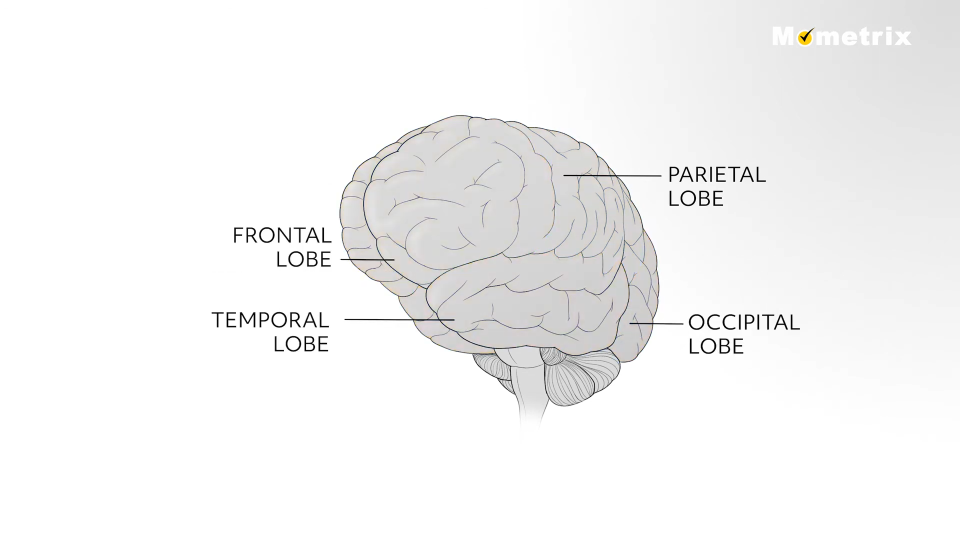
The parietal lobe, found on the “crown” of your head and directly posterior to the frontal lobe, is responsible for receiving and processing information of the senses. The occipital lobe is located at the back of the skull inferior to the parietal lobe. This lobe is responsible for the processing of visual information. The temporal lobe, found directly inferior to the parietal lobe and over the ear area, is involved in emotional responses, speech, auditory processing, and language comprehension.
Cerebellum
The cerebellum is the area of hindbrain that is located under the cerebrum. Its function is to control movement coordination, regulate muscle tone, and maintain balance and equilibrium. The cerebellum interprets sensory information from the brain and peripheral nervous system which allows it to maintain fine motor control while hindering involuntary movement.
Damage to the cerebellum can occur as a result of alcohol, drugs, strokes, head injuries, cancer, and degenerative diseases. With any of these detrimental ailments to the cerebellum, you can expect to see problems in individuals dealing with speech, muscle tone, balance, and coordination of movement.
Brainstem
The brainstem is the final pathway from the brain to the spinal cord and is responsible for many automatic functions. Heart rate, breathing, digestion, and even swallowing are maintained by the brainstem. As a result, damage to this area of the brain can be serious and sometimes fatal. Even if you survive an injury to the brainstem, there is still a high chance you could be in a vegetative state.
The brainstem is composed of the midbrain, pons, and medulla oblongata. Each area of the brainstem controls different functions.
For example, the pons is responsible for facial expressions and posture, whereas the midbrain is associated with temperature regulation and motor control. The medulla has the most important roles dealing with the involuntary functions such as breathing, heart rate, and blood pressure.
Brain Matter
The brain is also composed of two types of tissue- grey matter and white matter. The grey matter, named for its pinkish-grey color, is where the cell bodies, dendrites, and axon terminals of neurons reside. This is the area where synapses between neurons occur. This tissue is found throughout the cerebellum, cerebrum, and the brain stem.
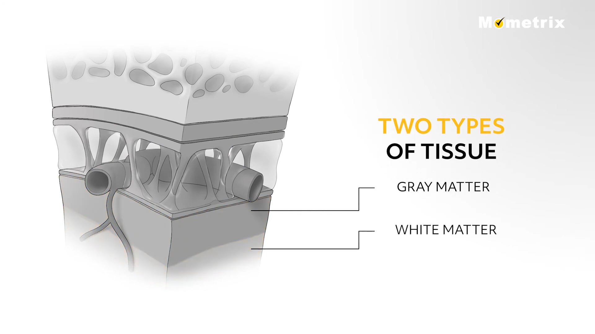
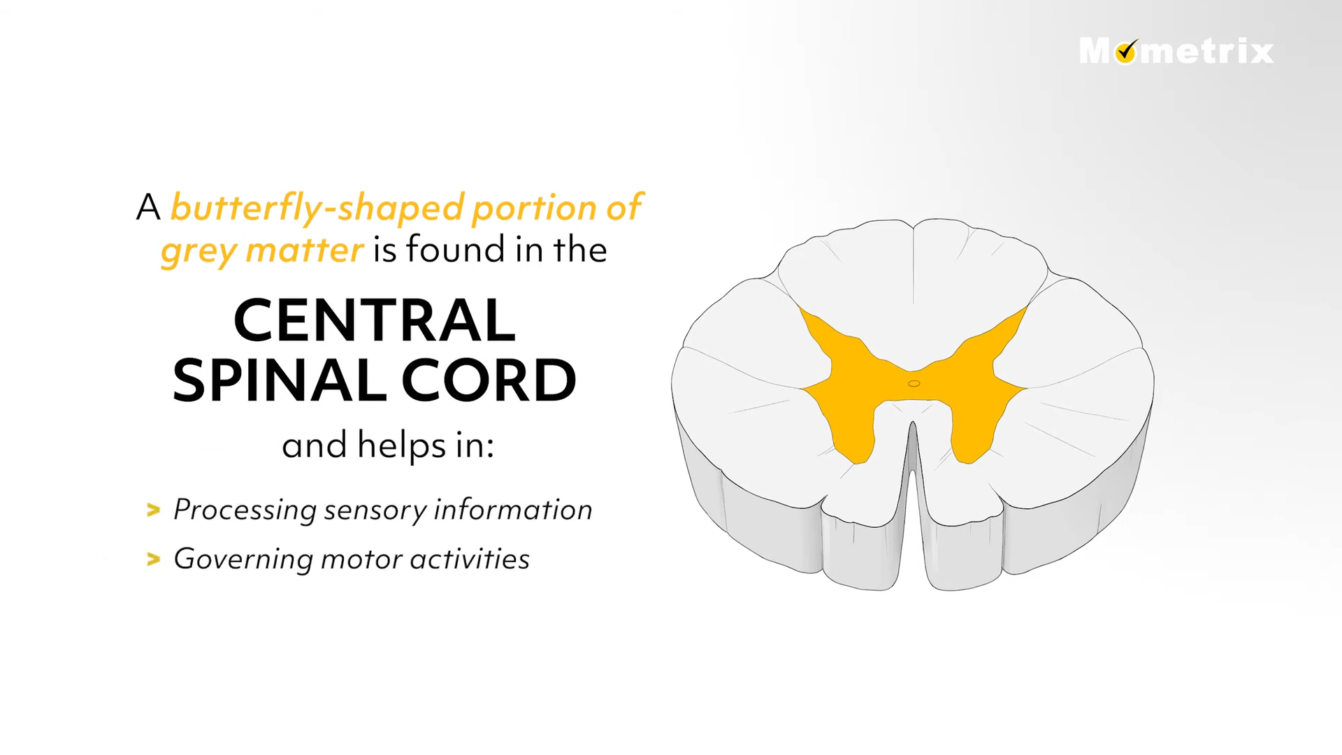
A butterfly-shaped portion of grey matter is found in the central spinal cord and helps in processing sensory information and governing motor activities. The white matter of the brain lays under the gray matter and is composed of nerve cell axons. The function of the white matter is to conduct and send nerve signals throughout the spinal cord. Damage to the white matter of the brain can cause delayed reflexes, sensory problems, and movement disorders.
Review Questions
Let’s test our knowledge using our brain!
1. Upon reviewing the MRI of an elderly patient who suffered a stroke to the right hemisphere of the brain, you can expect:
- Weakness to the left side of the body
- Weakness to the right side of the body
- Weakness to both sides of the body
The brain is divided into two hemispheres which control opposite sides of the body, therefore a stroke in the right hemisphere of the brain will cause weakness in the left side of the body.
2. A patient comes in to the emergency room and is complaining of visual disturbances and inability to focus. Which area of the brain is probably affected?
- Parietal
- Frontal
- Occipital
- Temporal
The occipital lobe of the brain is responsible for processing visual information.
By knowing the parts of the brain and their associated functions, you can anticipate the damage one might suffer following a brain injury.
Thank you for watching this brief video over the anatomy of the brain!
