

Hey guys welcome to this Mometrix video over cell structure!
Cells are foundational to life. In 1839 Matthias Schleiden and Theodor Schwann formulated the cell theory. The cell theory historically contains three sub-theorems:
- Every living thing is made up of a cell or multiple cells
- The cell is the basic unit of life
- Cells come about via cell division.
So, there must be an already existing cell for a new cell to come about.
The modern version of cell theory has added three more sub-theorems to the existing three, making six:
- Energy transference from one cell to another happens from within a cell
- All hereditary information is located within cells
- All cells contain the same basic chemical composition
So, basically what cell theory tells us is that cells are pretty important, and understanding the structure of cells, the most basic unit of life, is crucial to understanding the rest of an organisms make-up and function.
In this video we will look at the main components of eukaryotic animal cell structure: cell membrane, nucleus and nucleolus, cytoplasm, and cytoplasmic organelles.
Cell Membrane
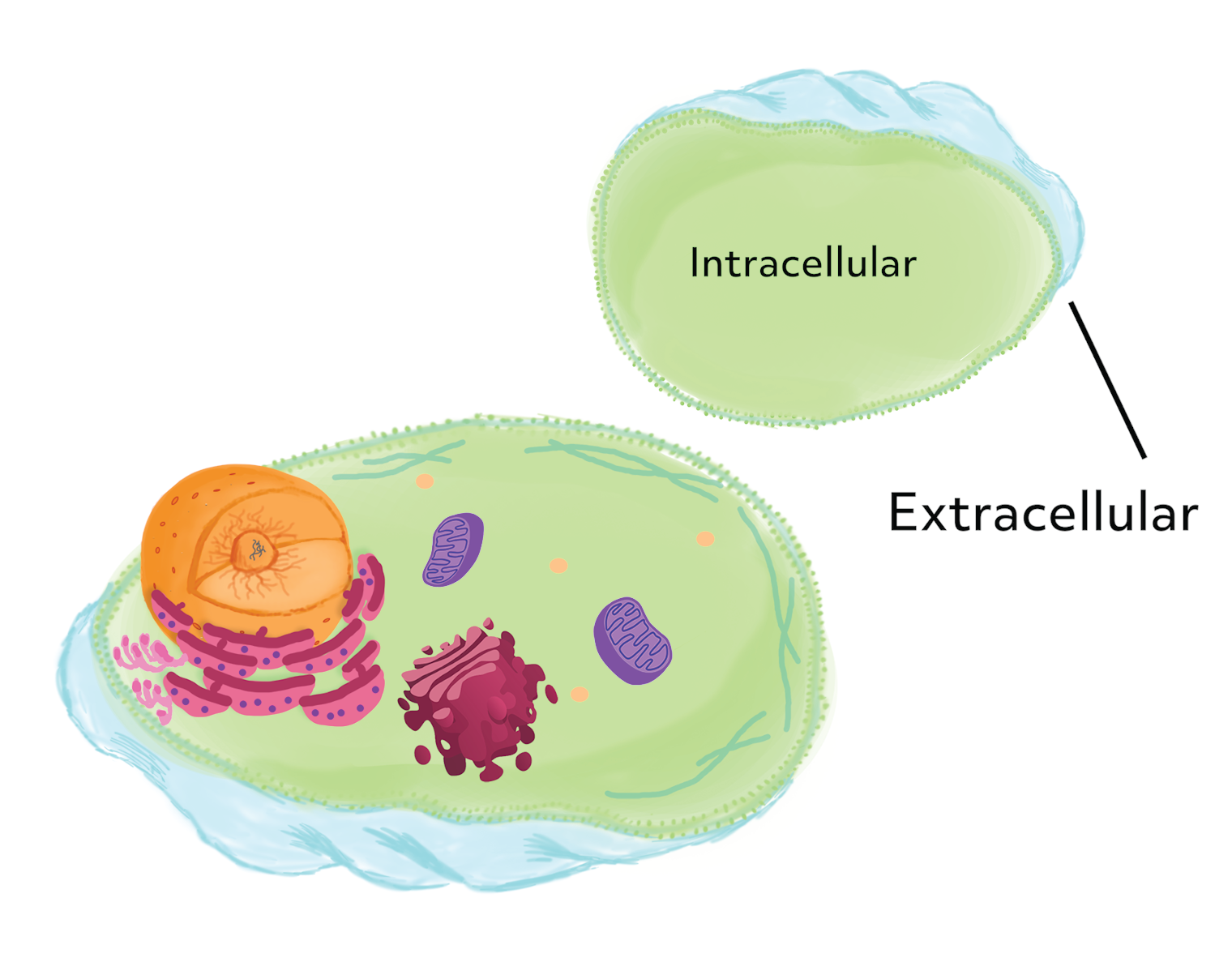
You have a bunch of stuff inside of a cell, the material on the inside is referred to as intercellular, and you have a bunch of stuff outside of a cell, this material is called extracellular.
Well, the cell membrane is what separates the extracellular material from all the intercellular material. The cell membrane maintains the coherence of the cell, and controls what passes in and out of the cell.
The cell membrane is made up of a double layer of phospholipids and proteins that control the transporting of substances in and out of the cell.
Located within the cell membrane is the nucleus. The nucleus is the most prominent structure within the cell membrane. The outermost portion of the nucleus is also made up of double layer phospholipids and proteins called the nuclear envelope (or outer and inner nuclear membrane). On the innermost surface of the inner nuclear membrane is a stabilizing network of fibrils called the nuclear lamina. The nuclear lamina helps to give structure and stability to the nuclear membrane.
Interspersed on the outside of the nuclear envelope are small pores called nuclear pores, which regulate the passage of molecules, ions, and RNA. Within the nucleus is a semi-solid fluid called nucleoplasm. Suspended in the nucleoplasm is the nucleolus and chromatin. The nucleolus take up about a quarter of the nucleus’ volume; it is made up of proteins and ribonucleic acid (or RNA).
Chromatin is a long strand of DNA, made up of numerous genes. Chromatin forms chromatids, and two chromatids form a single chromosome. All of a cell’s chromosomes are located within the nucleus. What you will also see dispersed throughout the nucleus are little particles called ribosomes, which are made in the nucleolus, and being transported out of the nucleus through the nuclear pores and into the cytoplasm.

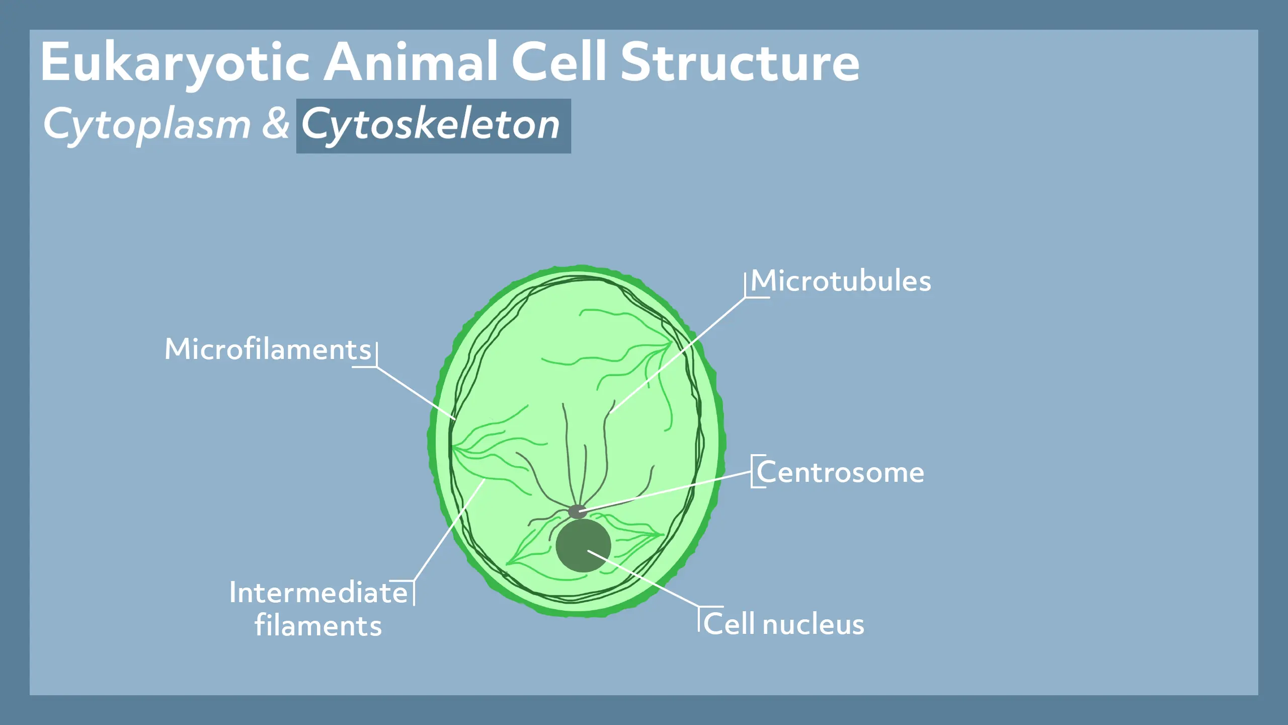
Cytoplasm and Cytoskeleton
Cytoplasm is a gel-like fluid that fills the entire inside of a cell, and suspends all of the cell’s inner components. The cytoplasm is composed primarily of water and salt. It plays a major role in giving the cell its shape. Without the cytoplasm the cell would be deflated, and cellular activity would not be possible.
The cytoskeleton is a component in the cell that does exactly what it sounds like. It gives the cell structure, shape, and stability, and allows for movement just like our skeletal system does for us. The cytoskeleton is comprised of three main materials: microfilaments, intermediate filaments, and microtubules. Microfilaments are, like their name suggests, tiny, tiny, filaments that look like thin threads. Intermediate filaments are a little larger than microfilaments, and the largest of the three are the microtubules. Microtubules look like microscopic size tubes.
Cytoplasmic Organelles
Cytoplasmic organelles are often referred to as “little organs,” which is what organelle means. They are structures bound within the membrane of the cell. Cytoplasmic organelles, other than the nucleus, are mitochondria, ribosomes, endoplasmic reticulum, golgi apparatus, and lysosomes.
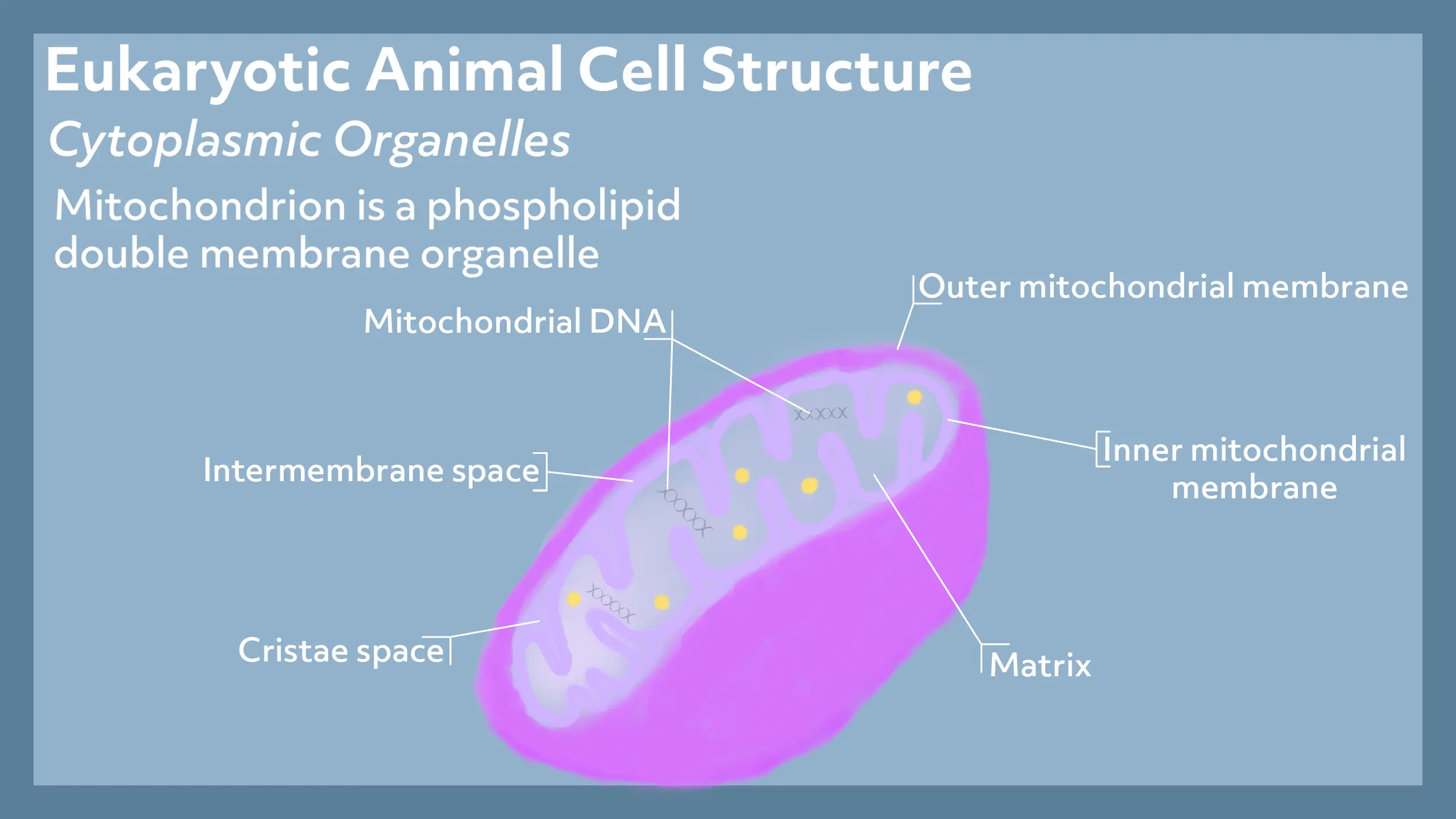
A mitochondrion is also a phospholipid double membrane organelle, and is shaped sort of like a bean. The mitochondrion has five major chambers or compartments: the outer mitochondrial membrane, the inner mitochondrial membrane, the intermembrane space (which is the space between the outer and inner mitochondrial membrane), the cristae space (which is the space created between the inner membrane foldings), and the matrix (which is all the space on the inside of the inner mitochondrial membrane). A mitochondrion also contains its own DNA called mitochondrial DNA, which is found within the matrix of a mitochondrion.
Ribosomes are tiny particles located all throughout the cytoplasm of a living cell. They are made up of 60 percent ribosomal proteins, and 40 percent protein molecules. A ribosome has two subunits: the large subunit, and the small subunit.
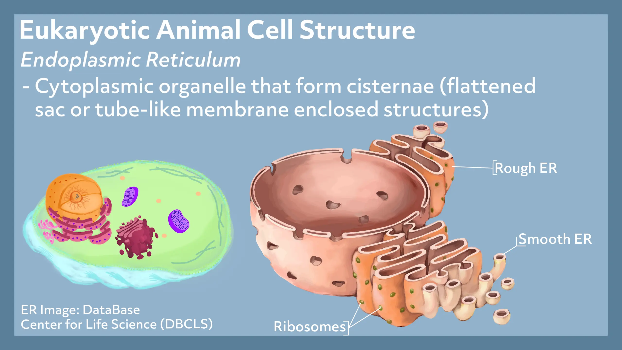
Endoplasmic Reticulum
The endoplasmic reticulum (or ER) is a cytoplasmic organelle that form cisternae (which are flattened sac or tube-like membrane enclosed structures). The ER is attached to the nuclear envelope of the nucleus. There are actually two types of ER: rough ER, and smooth ER. Rough ER looks like sheets or sacs of rugged membranes. Smooth ER look like slick tubes. The reason rough ER looks rough is due to the ribosomes that are attached to the outside. Smooth ER does not have ribosomes attached to it. Most cells contain ER, except for red blood cells and sperm cells. The size of the ER, in the cells that do have ER, varies based on the function of that cell.
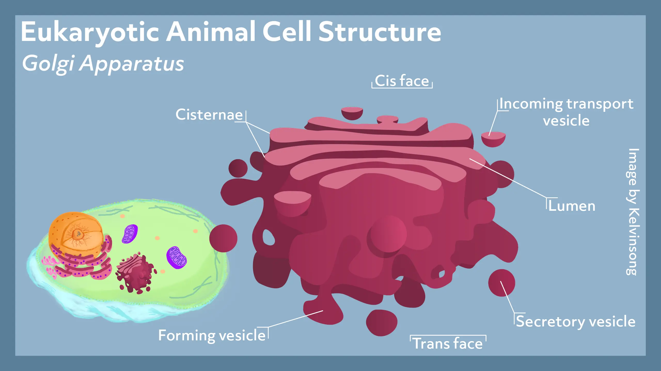
Golgi Apparatus
The Golgi apparatus is a cytoplasmic organelle made up of cisternae. The pockets within the cisternae are referred to as the lumen. The Golgi apparatus sits in the cytoplasm near the rough ER. The Golgi apparatus has two faces: the Cis face and the trans face. The cis face is the receiving side of the Golgi apparatus, and the trans face is the secreting side of the Golgi apparatus. The cis face is the side closest to the rough ER, and it receives the vesicles from the rough ER. The trans face secretes those vesicles.
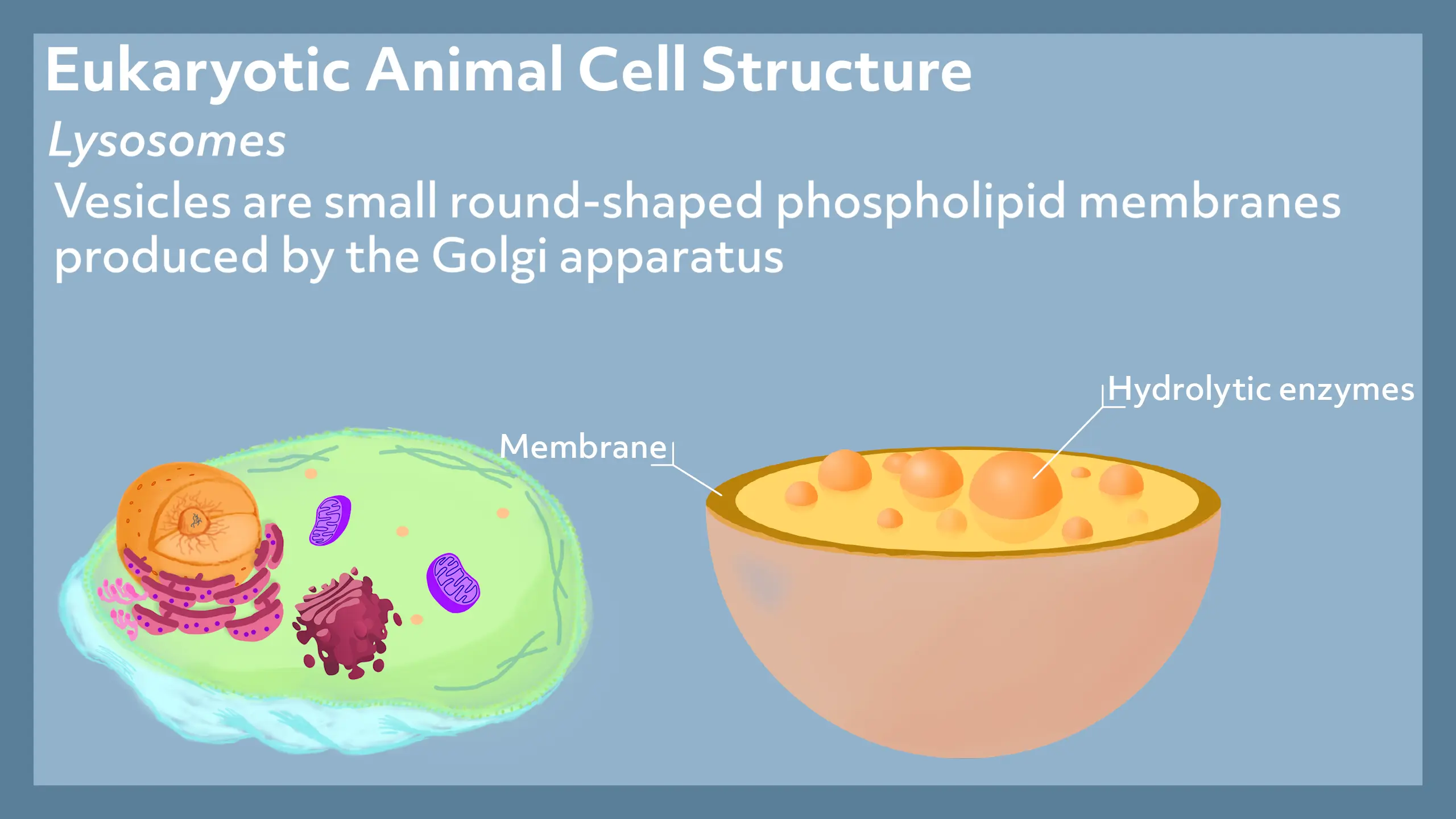
Lysosomes
Vesicles are small round shaped phospholipid membranes produced by the Golgi apparatus. Within the membrane of a lysosome acidity is maintained by a proton pump. Digestive enzymes are also contained within lysosomes.
So, that is an overview of the structure of a eukaryotic animal cell.
See you guys next time!