
Welcome to this video on the cardiovascular system! The cardiovascular system, also known as the circulatory system, is made up of the heart, blood vessels, and blood that circulates throughout the body. You will also hear the term the ‘cardiopulmonary system,’ which is referring to the cardiovascular system along with the trachea, bronchi, and lungs (the pulmonary portion).
In this video, we will take a closer look at the cardiovascular system: the heart’s basic anatomy, two types of circulation, how blood circulates through the heart, what causes the heart to beat, and the makeup of blood.
Heart’s Anatomy and Function
The heart is a muscle that sits in the upper left side of the chest behind the rib cage. It weighs roughly 10 ounces and is about the size of a clenched fist. The heart is made up of a system of chambers and valves; the chambers momentarily hold blood while the valves allow blood to flow in one direction to the next chamber. It beats about 100,000 times a day and sends blood throughout some 60,000 miles of blood vessels. There are several components that help the heart function properly.
The heart is composed of three layers: the epicardium, the myocardium, and the endocardium. The epicardium forms the outermost layer of the heart wall and protects the heart’s inner layers. It contains the coronary blood vessels which supply blood to the heart. The myocardium layer is like a pump, contracting to pump blood out of the heart and then relaxing to let the heart fill with blood again. The endocardium, the third layer of the heart, is a thin inner layer that lines the heart’s chambers. Let’s take a look now at the internal anatomy of the heart.
The heart is divided up into four chambers: the right atrium and right ventricle, and the left atrium and left ventricle. When we refer to the right side of the heart, we’re referring to the side of the heart in relation to the right side of a person’s body. The left side of the heart is on the left side of the body.
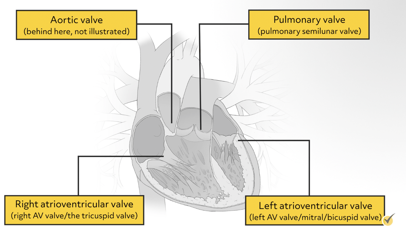
The heart also has four one-way valves:
- the right atrioventricular valve, also called the right AV valve or the tricuspid valve
- the pulmonary valve or the pulmonary semilunar valve
- the aortic valve, and
- the left atrioventricular valve, also called the left AV valve or the mitral/bicuspid valve.
One-way, or unidirectional flow is important. If the valves are leaking and allow blood to flow in both directions, then the heart has to work harder to pump the same amount of blood. This is called valve regurgitation.
Now that we’ve looked at the chambers and valves of the heart, let’s talk about the blood vessels that connect everything and allow for the proper functioning of the heart.
Veins and Arteries
As a rule, veins bring blood towards the heart, and arteries, which starts with an “a,” carry blood away from the heart.
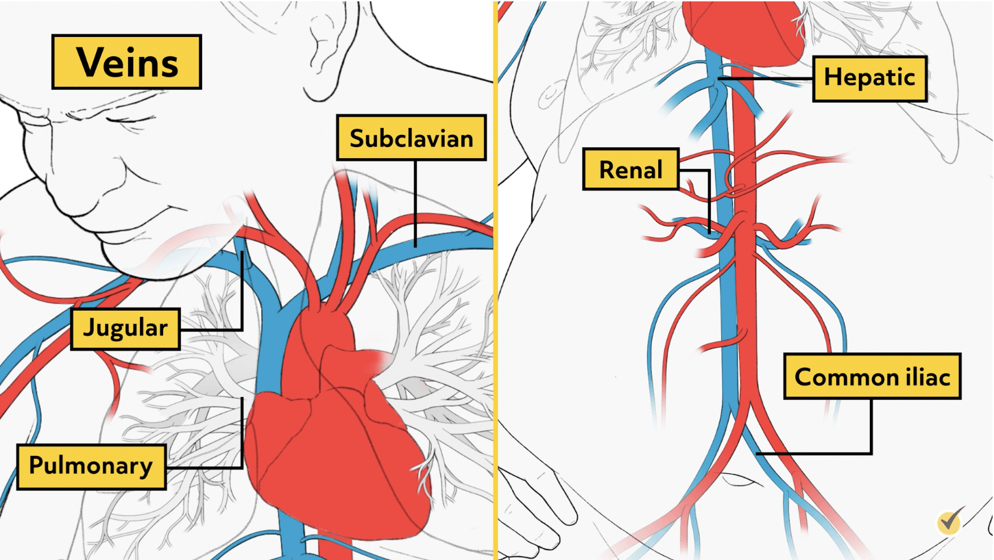
The major veins that bring blood to the heart are the superior vena cava and the inferior vena cava. The superior vena cava brings blood to the heart from the upper body while the inferior vena cava brings blood from the lower body. Other veins in the heart include the right pulmonary veins and the left pulmonary veins. The right and left pulmonary veins bring oxygenated blood from the lungs into the heart. Other major veins include the jugular vein, left and right subclavian veins, hepatic portal, renal veins, and the common iliac veins. Moving further from the heart and closer to the extremities, the blood vessels get smaller and the veins become venules.
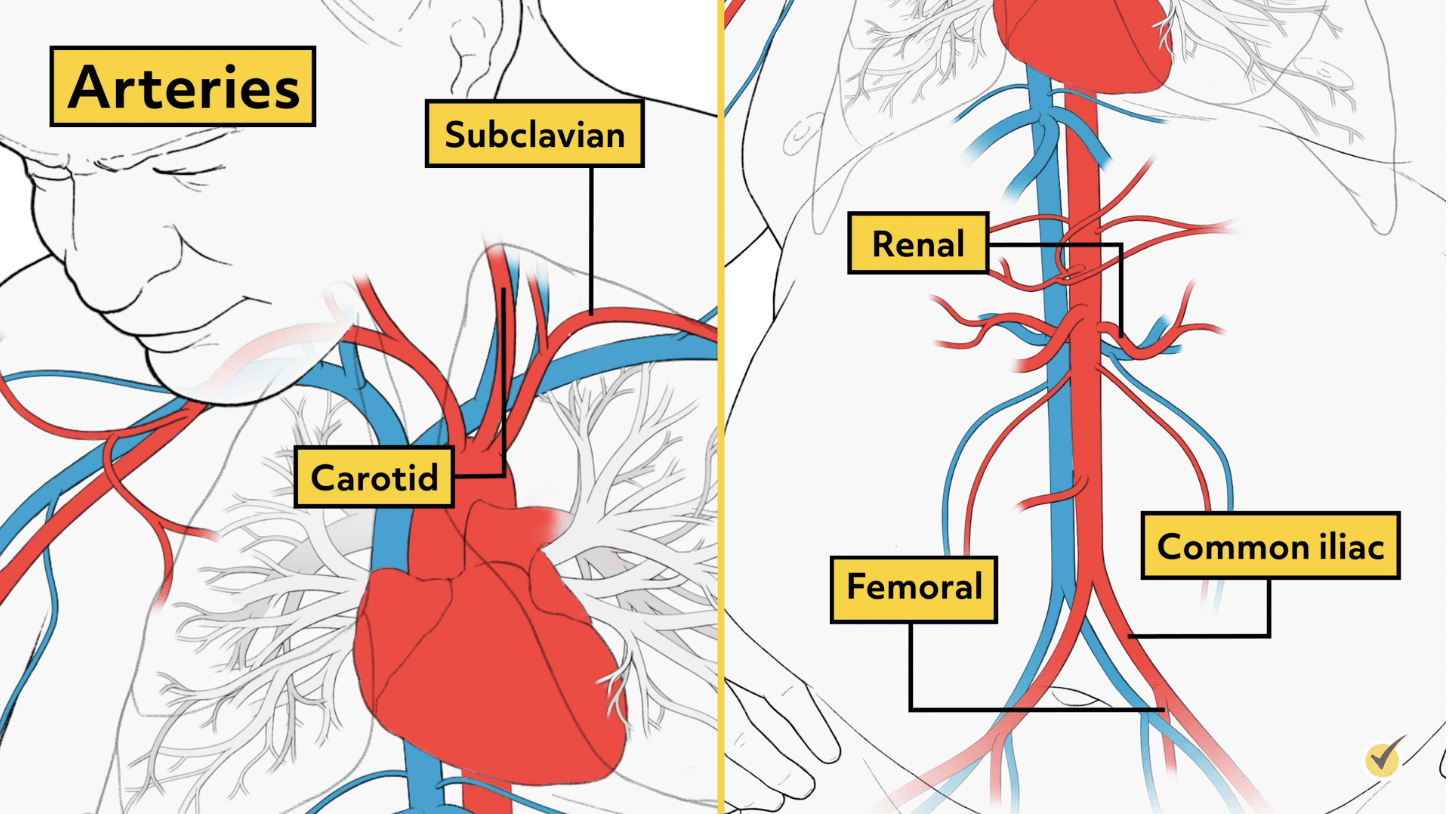
The heart also has arteries, taking blood away from the heart. Some major arteries include:
- the right pulmonary artery,
- the left pulmonary artery,
- and the aorta.
The aorta branches off into smaller arteries and is responsible for distributing blood throughout the body once it’s been oxygenated, so this is the biggest, strongest artery in the body. The left and right pulmonary arteries carry deoxygenated blood to the lungs, where the blood gets oxygenated. The heart also has coronary arteries that wrap around the outside of the heart and supply blood to the heart muscle. They are called the left coronary artery and the right coronary artery. Other major arteries in the body include the carotid artery, subclavian artery, common iliac artery, femoral artery, and renal artery. As we move further from the heart, these arteries become arterioles, and arterioles become very fine capillaries.
Arteries and veins are connected by way of microscopic blood vessels called capillaries that make up a capillary bed. Capillary beds are a network of capillaries located between arterioles and venules where nutrients and wastes are exchanged.
Blood Circulation
Now let’s look at the circulation of the blood through the body. There are two types of circulation, pulmonary circulation, where blood moves between the heart and the lungs; and systemic circulation, which is the path blood takes between the heart and all organs and tissues in the rest of the body. Pulmonary circulation transports deoxygenated blood to the lungs to absorb oxygen and release carbon dioxide. The oxygenated blood then flows back to the heart, where it gets pumped out to the systemic circulation (the rest of the body).
Blood Traveling Through the Heart
Let’s take a closer look at the pathway of blood as it travels through the heart. Starting with oxygen-poor blood in the superior vena cava, this blood will flow into the right atrium where it is then forced through the right AV valve into the right ventricle. Blood passes through the pulmonary semilunar valve and is forced up into the pulmonary arteries so that it can be oxygenated in the lungs.
Once the blood is oxygenated, it reenters the heart through the right and left pulmonary veins and travels into the left atrium. From here, the blood is forced through the left AV valve and into the left ventricle. Finally, the blood travels through the aortic valve and into the aorta where it can be distributed to organs and tissues throughout the body that need oxygen most. The real gas exchange takes place in the capillaries where tissues trade their carbon dioxide for oxygen the blood is carrying. Now that the blood has given its oxygen to the tissues, it returns by way of veins into the superior and inferior vena cava where we started so that it can flow back through the heart and repeat the process.
Cardiac Cycle
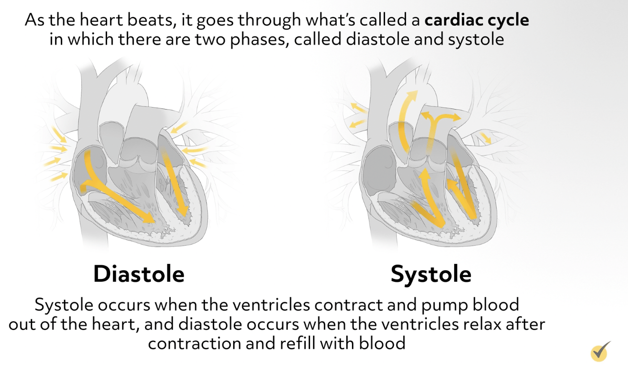
As the heart beats, it goes through what’s called a cardiac cycle in which there are two phases, called diastole and systole. Systole occurs when the ventricles contract and pump blood out of the heart, and diastole occurs when the ventricles relax after contraction and refill with blood. This systolic and diastolic process measures blood pressure. Systole (or the top number) is basically the pressure required to pump blood out of the heart; and diastole (the bottom number) is the pressure that remains when the heart is in a resting state between heartbeats.
You may be wondering what causes the heart to beat. Electrical impulses from the heart muscle (the myocardium) cause the heart to beat (contract). Known as the cardiac conduction system, a group of specialized cardiac muscle cells in the walls of the heart send signals to the myocardium causing it to contract. The main components of the cardiac conduction system are the SA node, AV node, bundle of His, bundle branches, and Purkinje fibers. The sinoatrial (SA) node, located at the top of the right atrium, starts the sequence by causing the atria to contract. The SA node is referred to as the ‘natural pacemaker.’ From there, the signal travels to the atrioventricular (AV) node, through the bundle of His, down bundle branches, and through Purkinje fibers, causing the ventricles to contract. These signals of electrical current can be seen on a graph called an electrocardiogram (EKG).
Blood Composition
Now we’ll take a quick look at the red fluid that flows through our cardiovascular system. Blood has four main components – plasma, red blood cells, white blood cells, and platelets.
The blood that runs through the veins, arteries, and capillaries is known as whole blood. Whole blood contains about 55% plasma and 45% blood cells. The average man has about 12 pints of blood and the average-size woman has about 9 pints.
The liquid component of blood is called plasma, which transports blood cells, nutrients, waste products, antibodies, clotting proteins, and hormones throughout the body.
Red blood cells contain the protein hemoglobin that carries oxygen from the lungs to the rest of the body and then returns carbon dioxide from the body to the lungs to be exhaled. Red blood cells are made inside our bones and get their red color from the hemoglobin.
White blood cells are the fighters because they protect our bodies against infection.
Platelets are actually small fragments of cells that form small clots to help the body stop bleeding.
Blood has several different functions including transporting oxygen and nutrients to the organs and tissues in our body, carrying cells and antibodies that fight infection, taking waste products to the kidneys and liver so they can filter and clean the blood, and forming blood clots to stop excess bleeding.
Now that you’re familiar with the heart’s anatomy and the path blood takes as it circulates through the heart and body, let’s look at this quick review question:
Review Question
Based on what you’ve learned about the circulation of blood through the heart and lungs, which of the following vessel(s) would contain oxygenated blood?
- Inferior vena cava
- Right and left pulmonary arteries
- Right and left pulmonary veins
- Aorta
- Both C and D
The pulmonary veins and the aorta. The inferior vena cava brings deoxygenated blood from the lower body to the heart and the pulmonary arteries are also carrying deoxygenated blood to the lungs to get oxygenated. Blood in the right and left pulmonary veins just came from being oxygenated in the lungs and the aorta is carrying that oxygenated blood from the left ventricle to the rest of the body.
I hope this review was helpful and happy studying!
- “Circulatory System Anatomy, Diagram & Function.” n.d. Healthline
- Toro, Ross. 2013. “Diagram of the Human Circulatory System (Infographic).” Live Science
- Bailey, Regina. 2016. “The Layers of the Heart Wall.” ThoughtCo
- “Conditions and Diseases.” n.d.
- Healthwise. 2019. “How the Heart Works.” Alberta.ca
- “Left Atrium Function, Definition & Anatomy | Body Maps.” 2018. Healthline.
- Lumen Learning. 2019. “Circulation and Heart Valves | Boundless Anatomy and Physiology.”