

Welcome to this video on the anatomical and clinical parts of teeth.
Types of Teeth
An infant’s primary teeth first begin to erupt at about 6 months of age on average. These teeth are commonly referred to as the “baby teeth” and begin to shed when the child is about 6 years old, when they are replaced by permanent teeth.
By about age 11, most children have shed all of their primary teeth and have their permanent dentition, or set of teeth, although the last 4 molars usually don’t come in until about age 21.
These last molars are called “wisdom teeth” and are often removed because they become impacted, which means they did not have the room they needed to emerge properly.
While the primary teeth consist of only 20 teeth, a full set of permanent teeth consists of 32 teeth or 28 if all four wisdom teeth are removed. There are four different types of teeth:
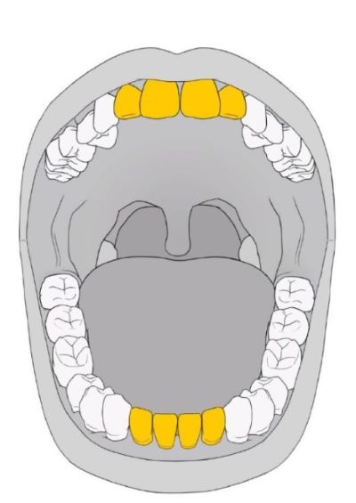
- Incisors
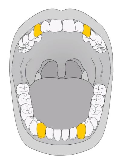
- Canines, also known as cuspids
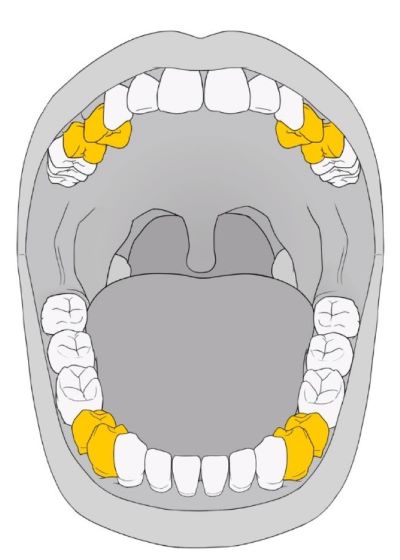
- Premolars, also known as bicuspids AND
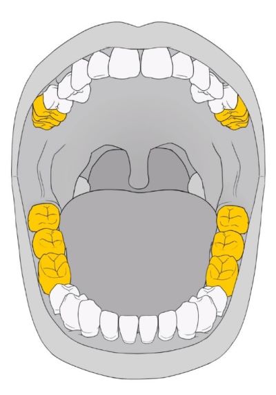
- Molars
There are eight incisors, four in the upper jaw and four in the lower. These are what we usually call our front teeth and are intended to help you bite into food. Incisors are also the first teeth that erupt in infants.
The four canine teeth are the sharp, pointed teeth on either side of the incisors, top and bottom. They aid in the process of tearing food.
There are 8 premolars or bicuspids, two behind each canine tooth, top and bottom. They have flat ridged surfaces and help crush and grind food.
The last teeth are the 12 molars, three behind the premolars, top and bottom, and these include the wisdom teeth. These teeth are the largest and help to grind food into small enough pierces to easily swallow.
Anatomy of Teeth
While the different types of teeth may have a different shape and appearance, the basic structure of all teeth remains the same. The parts of a tooth include the
- Crown
- Enamel
- Dentin
- Pulp
- Root
- Cementum AND
- Periodontium
The crown is the part of the tooth that we see if we look into the mouth. The anatomic crown extends to below the margin of the gum while the clinical crown is just the visible part of the tooth. The anatomic crown always has the same size, but the clinical crown may vary in size.
For example, the clinical crown may enlarge if the gums recede and expose more of the tooth. The clinical crown may also decrease in size if the gums are inflamed and swollen.
The entire crown is covered and protected by a layer of enamel. Enamel is white, although some may have a yellow or gray tinge. Enamel is the hardest material found in the body, able to withstand a force of 100,000 pounds per square inch, though it is still susceptible to cracking or chipping.
The next layer of the tooth is the dentin, which makes up the major portion of the tooth. Dentin extends from the crown to the roots and comprises somewhat elastic mineralized tissue that is harder than bone, but not as hard as enamel.
Dentinal fibers are found within dentin, and these fibers can transmit pain and sensations of heat and cold, so dentin is considered living tissue.
The inner surface of the dentin serves as the walls of the pulp cavity. In the crown, the dentin is covered by enamel, but as it extends into the roots of the tooth, the dentin is covered by cementum.
The pulp is the inner layer of the tooth and contains blood vessels and nerves, so this part of the tooth is living. The part of the pulp that is found in the crown is called the coronal pulp and the part in the roots is the radicular or root pulp. Pulp helps to form dentin.
Once a tooth is mature, it no longer requires pulp and can survive, as long as the pulp is not infected.
The root is the part of the tooth that is anchored in the alveolar process, which is the part of the jawbone that contains the tooth socket. The tapered tip is called the apex of the root. Roots may be
- Singular, such as those found in incisors and canines,
- Bifurcated (divided into two parts), such as those commonly found in the mandibular premolars, OR
- Trifurcated (divided into three parts), such as those found in the maxillary molars.
Remember that while the crown is covered by enamel, the roots are covered by cementum. The point at which the enamel of the crown becomes the cementum of the root is called the cervical line or the cementoenamel junction.
The periodontium provides protection, support, and nourishment to the tooth. The periodontium comprises the gum, the cementum, the alveolar bone or process, and periodontal ligaments.
The periodontal ligaments connect the cementum to the alveolar bone in the tooth socket.
Cementum is rigid connective tissue that has a yellowish color. It can sometimes be seen at the base of a tooth if the person has periodontal disease and receding gumlines.
That’s all for now – thanks for watching and happy chewing.
- “Anatomic Crown: What It Is & the 3 Layers | Colgate®.” n.d.
- Hoffman, Matthew. 2009. “Picture of the Teeth.” WebMD
- “Tooth Anatomy – Gosford, Experienced Dentists.” VC Dental. 2019
- Watson, Stephanie. 2018. “What Are the Different Types of Teeth Called?” Healthline
- “Parts of the Tooth | Complete Anatomy.” 2021