BEST Mnemonics for Heart Anatomy and Physiology

Welcome to this video tutorial on heart mnemonics. First of all, we’re going to take a look at the anatomy of the heart.
Heart Anatomy
Each half of the heart has an upper collecting chamber, the atrium, and a lower pumping chamber, the ventricle.
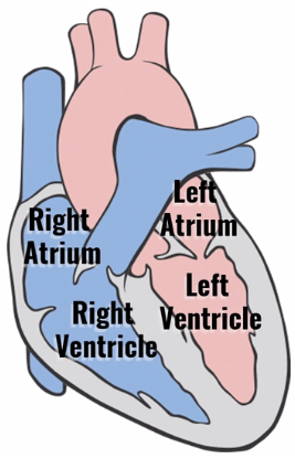
You can remember their location because A comes before V. The atrium is above the ventricle.
Heart Sounds
Next, we’ll take a look at the heart sounds heard with a stethoscope. As blood flows from the atria to the ventricles, it passes through the tricuspid and bicuspid valves, also known as the atrial ventricular or AV valves. These valves then snap shut, making the first heart sound S1, or “lub” in “lub dub.”
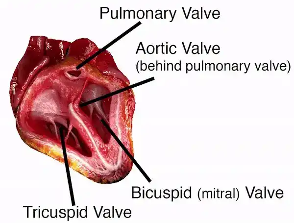
As these two valves snap shut, the pulmonary and aortic valve just opened. This is the beginning of systole or contraction of the ventricles. As the ventricles contract, blood is pumped through the pulmonary valve leading to the lungs and the aortic valve leading to the aorta and the rest of the body.
These valves then snap shut making the second heart sound S2 or “dub” in “lub dub.” This is the end of systole and the beginning of diastole. At the same time, the tricuspid and bicuspid valves just opened and the process repeats.
Blood Flow
Blood flows from the right atrium through the tricuspid valve to the right ventricle, then on through the pulmonary valve to the pulmonary artery and lungs.
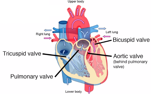
Pulmonary veins return blood to the left atrium through the bicuspid, or mitral, valve to the left ventricle and through the aortic valve, to the aorta and the rest of the body. Sound confusing? Just remember this mnemonic for the order of blood flow through the valves:
Try
Performing
Better
Always(Tricuspid, Pulmonary, Bicuspid, and Aortic.)
Stethoscope Placement for Heart Sounds
Listening to the heart is accomplished by using five systematic locations on the anterior chest wall.
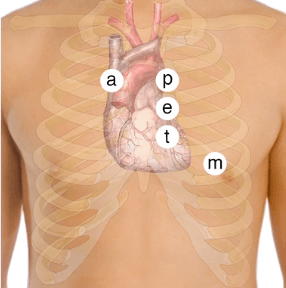
These landmarks are the places on the chest where you can best hear sounds from each of the respective heart valves, even though they are not at the exact anatomic site.
The stethoscope is positioned downstream from the flow of blood through the valves. Use the diaphragm of the stethoscope switching to the bell to hear lower-pitched sounds. The aortic valve area is on the right side at the second intercostal space. The pulmonic valve area is located on the left side at the second intercostal space.
Erb’s point is at the left third intercostal space. The tricuspid valve area is at the left fourth intercostal space, and the mitral valve area is at the left fifth intercostal space, at the mid-clavicular line. You can remember these locations by remembering this mnemonic:
All
People
Enjoy
Time
Magazine(Aortic, Pulmonic, Erb’s point, Tricuspid, Mitral)
EKG Placement
When placing EKG leads on a patient, proper skin preparation and lead placement are essential for a clear EKG reading. Start by cleaning the skin briskly with an alcohol rub and allow to dry. Place the electrodes in their proper location and press firmly.
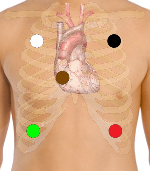
For placement, remember “white on right, with white clouds over green grass.” On the left, “smoke over fire (or black over red) with brown in the middle.”
Heart Murmurs
Heart murmurs are audible vibrations of the heart and major vessels that occur due to turbulent blood flow. Murmurs are caused by abnormal blood flow or a structural alteration in the heart or the walls of the major vessels.
Some murmurs are innocent and may disappear over time or last a lifetime without causing further health problems. Other murmurs are abnormal and more serious. You can remember the various causes of heart murmurs by remembering SPAMS.
Stenosis of a valve
Partial obstruction
Aneurysms
Mitral or aortic regurgitation
Septal defect
S for stenosis, or narrowing of a valve. P for partial obstruction, which may be due to calcification, valve degeneration, coronary artery disease, or congenital abnormalities. A for aneurysms, in which an area of the aorta has become weak and enlarged. M for mitral or aortic regurgitation. This is when blood is leaking backward through the mitral or aortic valve. The heart has to work harder to force blood through the damaged valve and over time this can weaken or enlarge the heart leading to heart failure. S for septal defect, this is a hole in the septum that separates the ventricles or the atria.
Heart Blocks
A heart block is an abnormal heart rhythm known as an arrhythmia and can occur anywhere in the specialized conduction system of the heart. The electrical signals telling the heart to contract are partially or totally blocked between the atria and ventricles. Therefore, it is called an atrioventricular, or AV block.
The AV blocks are identified by their severity: first-degree or second-degree, which can be subdivided into Type I (Wenckebach) and Type II (Mobitz II), and then third-degree heart block. What does all of this mean? Here’s our further explanation.
First-Degree
First-degree heart block occurs when the electrical impulses are slowed down but they all successfully reach the ventricles.
This type rarely causes problems and is often seen in highly trained athletes, or as a result of some medications. On the EKG, the PR interval is greater than 0.2 seconds, indicating a conduction delay in the AV node.
Second-Degree
Second-degree heart block exists when there is intermittent conduction failure and occurs in two varieties.
Type I (Wenckebach)
In Wenckebach, the electrical impulses are delayed further and further until conduction fails to reach the ventricles entirely. This is characterized by a PR interval that progressively lengthens until the P wave is not followed by a QRS complex—the QRS has dropped off after the P wave.
Type II (Mobitz II)
In this type, some of the electrical impulses are unable to reach the ventricles at all. The non-conducted P waves can’t get through and may occur at random or in patterned ratios. This is much more serious and may require a pacemaker.
Third-Degree
Third-degree heart block is a complete heart block in which none of the electrical impulses from the atria reach the ventricles, and the atria and ventricles beat independently. The ventricles may generate some impulses on their own but at a rate much slower than normal.
Generally, the patient requires a permanent pacemaker. Here’s a simple poem to help you remember the types of AV heart blocks.
If the R is far from P, then you have a first-degree
Longer, longer, longer drop, then you have a Wenckebach
If some Ps don’t get through, then you have Mobitz II
If Ps and Qs don’t agree, then you have a third-degree
Frequently Asked Questions
Q
How do I remember the order of blood flow through the heart valves?
A
Blood flows from the right atrium through the tricuspid valve to the right ventricle, then on through the pulmonary valve to the pulmonary artery and lungs. Pulmonary veins return blood to the left atrium through the bicuspid, or mitral, valve to the left ventricle and through the aortic valve to the aorta and the rest of the body. To remember the order of blood flow through the valves, remember this mnemonic:
Try Performing Better Always (Tricuspid, Pulmonary, Bicuspid, Aortic)
Q
How do I remember where to put the stethoscope to hear the different lung sounds?
A
The stethoscope is positioned downstream from the flow of blood through the valves The aortic valve area is on the right side at the second intercostal space. The pulmonary valve area is located on the left side at the second intercostal space. Erb’s point is at the left third intercostal space. The tricuspid valve area is at the left fourth intercostal space, and the mitral valve area is at the left fifth intercostal space, at the mid-clavicular line. You can remember these locations by remembering this mnemonic:
All People Enjoy Time Magazine (Aortic, Pulmonary, Erb’s point, Tricuspid, Mitral)
Q
How do I remember where to place the EKG leads in a 5-lead EKG?
A
The 5-lead EKG requires placement of the leads in specific regions to capture an accurate result. Regardless of machine, these leads are white, black, brown, red, and green. To remember the placement of these leads, think of the following phrases. For the patient’s right side, “White on right with white clouds over green grass.” For the patient’s left side and middle, “Smoke (black) over fire (red) with brown in the middle.”
Q
How do I remember the most common causes of heart murmurs?
A
Murmurs indicate abnormal or turbulent blood flow through specific vessels or chambers of the heart. The most common causes of heart murmurs can be remembered with the mnemonic SPAMS, Stenosis of a valve, Partial obstruction, Aneurysm, Mitral or aortic regurgitation, and Septal defect.
Q
How do I remember the types of heart blocks?
A
In a first-degree heart block the PR interval is greater than 0.2 seconds with an otherwise normal rhythm. In a second-degree type I (Wenckebach) heart block, the PR interval progressively lengthens until the P wave is not followed by a QRS complex—the QRS has dropped off after the P wave. In a second-degree type II heart block (Mobitz II), conducted P waves can’t get through and may occur at random or in patterned ratios. In a third-degree heart block, none of the electrical impulses from the atria reach the ventricles, and the atria and ventricles beat independently. Remember these with the following poem:
If the R is far from P, then you have a 1st degree
Longer, longer, longer, drop, then you have a Wenckebach
If some Ps don’t get through, then you have a Mobitz II
If Ps and Qs don’t agree, then you have a 3rd degree